* In vivo test of animal module of suppressing malignant tumor
The chosen C57BL/6 mice were 20 grams in an average of weight. Then, divided into bellowed 2 groups, 10 mice (5 males, 5 females) of each group for experiment.
- The controlled group: Each one injected with LLC-1(Lewis lung carcinoma) for 5×10⁶ cells, 7 days later, daily fed to basal diet . Daily observed till the 30th day for anatomy.
- The NCI-1 treated group :Each one injected with LLC-1(Lewis lung carcinoma) for 5×10⁶ cells,7 days later,daily fed to 2 grams of the “NCI-1extract” by weight of per kg of C57BL/6 in basal diet. Daily observed till the 30th day for anatomy.
Removed and weighed the tumors of both groups to obtain the results: ( as shown in Fig. 14)
- The average weight of tumor of the controlled group was 9.55±0.05 grams on the 30th day.
- The average weight of tumor of the treated group(NCI-1 group) was 4.52±0.05 grams on the 30th day.
It proved NCI-1 effectively suppressed the malignant tumor, cancer.
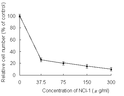
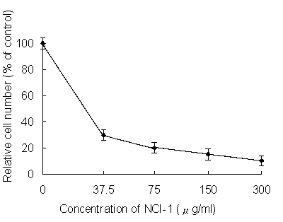
*For effects of tumor cytotoxicity by NCI-1(extract):
Summary:
| |
Cancer cell death
(at 300ug/ml of NCI-1) |
IC50
(ug/ml) |
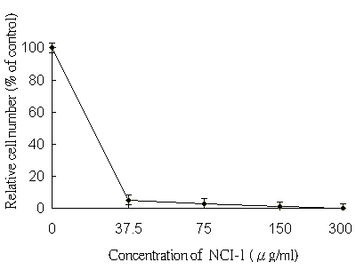
| Breast cancer |
up to 99% |
<37.5 |
| Lung cancer |
up to 99% |
37.5 |
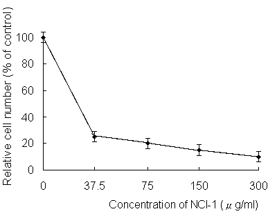
| Colorectal cancer |
up to 80% |
37.5 |
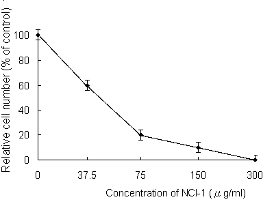
| Prostate cancer |
up to 70% |
75 |
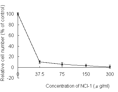
| Bladder cancer |
up to 99% |
<37.5 |
| Cervical cancer |
up to 80% |
<37.5 |
| Pancreas cancer |
up to 90% |
<37.5 |
| Gastric/Stomach cancer |
up to 90% |
<37.5 |
| Hepatoma/Liver cancer |
up to 90% |
<37.5 |
|
1. For MCF-7, cells of mammary gland of human breast adenocarcinoma:
2. For LLC-1, cells of Lewis lung carcinoma:
3. For SW480, cells of a primary adenocarcinoma of the human colon:

4. For Pc-3, cells of metastatic site of human prostate carcinoma:

5. For TSGH 8301, cells of human bladder carcinoma:
6. For Hela, epithelia cells of human cervical adenocarcinoma:

7. For Paca-2, cells of human pancreas carcinoma:
8. For AGS, cells of human gastric adenocarcinoma:
9. For HepaG2, cells of human hepatocellular adenocarcinoma:
* For in vivo bio-modification by NCI-1(extract):
1. NCI-1 for improving liver function of chronic hepatitis:

Figure 10 . It reduced more than 60% of abnormal AST/GOT.
2. The NCI-1 for improving liver function of acute hepatitis:

Figure 11. It reduced more than 55% of abnormal ALT/GPT.
3. The NCI-1 for improving platelets of hemostasis:
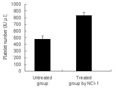
Figure 12. It overproduced 60% more of platelets in abnormal bleeding.
4. The NCI-1 for improving renal function:
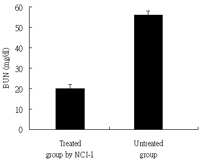
Figure 13. It reduced more than 64.7% of abnormal BUN.
5. The NCI-1 for treating cancers (in 30 days/animal module):
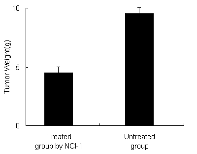
Figure 14. It suppressed 53% of malignant tumor.
-- The Animal Module of Cancer Therapy
The Animal Module of Cancer Therapy by Using NCI-1 (similar to early or middle stage of human cancer):
- C57BL/6 mice were divided to three groups, 10 mice/group (5 male, 5 female). One group was as the control group (Group 1), another one was fed daily 10mg as the low dose of NCI-1 (Group 2), the other one was fed daily 40mg as the high dose of NCI-1 (group 3). Inject Lewis lung carcinoma cell 4×10⁶/ml
- After 35 days and getting results: NCI-1 could suppress 85% of tumor mass growth
- The average tumor size of group 3 is 0.2cm3
- The average tumor size of group is 1.5cm3
-- Safety Evaluation/Treating Strategy
- 8 rats fed to 4g/rat of NCI-1 daily and 2 rats only fed to normal saline as the control group. After 90 day period, no toxicity or side effect was found
- In the clinical studies of NCI-1, some tumor markers was obviously reduced, including CEA, CA19-9, CA153, CA125, PSA/fPSA and AFP
- Tumors was obviously necrosis or disappeared examined by CT, MRI or ultrasonic examination
-- Scientific relations to Canavalia ensiformis (ConA) in NCI-1
- Con A receptors on the surface membrane of lymphocytes from patients with Hodgkins and other malignant lymphoma. - Proc.Natl.Acad.Sci.USA.1975
- Clinical and prognostic significance of Con A induced suppressor cell activity in malignant cervical neoplasia. - Bri.J.Obstet,Gynaecol.1990
- Con A induced supressor cells in relation to tumor growth and supressor T cell subset.-Int.J.Cancer.1981
- ConA induced supressor cell activity and autorosette forming cells in CML(leukemia)-Bri.J.Cancer 1983
- Interaction of ConA with the surface of virus-infected cell, - Cancer 1975
- Assessment of ConA reactivity of murine ascites tumor by inhibition of tumor cells migration.-Exper.Cell Res.1972
- Evidence for the presence of two cytokines in the supernant of ConA activated human pheral blood lymphocyte. - Annual meeting of the society for biological therapy.Pittsburg.USA.Nov.91.1991
- Lymphocytes exposed to ConA - J.of Cell.Phys.1990
- Differential toxicity on normal and transformed cells in vitro and inhibition of tumor development in vivo by ConA. - Nature.1970.
- Inhibitory effects of traditional Chinese medicines in agglutination assay with ConA,-J.Pharm.Soc.Japan.1992
- Combination therapy with 5-fluorouracil and L-canavaline(Canavalia ensiformis) in vitro and in vivo studies - Anticancer Drug,1995.
- ConA(from Canavalia ensiformis):potent mitogen used to stimulate cell proliferation in lymphocytes,primarily T-lymphocyte,culture. - National Institute Health.USA
- Cancer cells are readily aggregated by ConA,normal cells are not. Interaction of human plasma low density lipoprotein with ConA and with Ricin - J.Biol.chem.1975
- Differential effects of ConA and Suc –ConA on the macromolecular event of platelet activation.-Biochim.Biophys.Acta 1983
All the above statements are for educational purposes only
© 2006 National Cancer & Immune.LLC. All Rights Reserved
NCI-Con A 可減少鼻竇炎/鼻膿腫/氣喘/異位性皮膚炎
Marybeth S. Smuts/Laboratory of Developmental Biology and Anomalies, National Institute of Dental Research, National Institutes of Health, Bethesda, Maryland 20014
Concanavalin A (Con A), a lectin binding to mannosyl and glucosyl residues of glycoproteins and glycolipids, was used to study the appearance of carbohydrate-rich cell surface material on the olfactory placode and nasal processes which contribute to formation of the primary palate. In vivo incorporation of 3H-thymidine was also used in an attempt to correlate changes in labeling index with formation of the olfactory placode and nasal processes. The cell surface of the early front nasal epithelium binds Con A very little, if at all, but Con A binding was observed when the olfactory placode could be identified as a plate of cuboidal cells that exhibited a reduced labeling index. During the period of formation of the nasal processes, Con A binding was observed on the facial epithelium including the presumptive contact region. There was also a decline in the labeling index throughout primary palate formation.
NCI / Con A接合取下鼻腔發展中的複狀層疉物(息肉) ….Con A 結合鼻腔/嗅覺位置,可辨識出複合層疊細胞以減少沾黏指數…也減少形成主要味覺之沾黏指數 。
(1976年美國國家衛生研究院發表) (NCI-詳洽醫護營養人員)
__________________________________________________________
Deficient Concanavalin-A-induced suppressor-cell activity in patients with bronchial asthma, allergic rhinitis and atopic dermatitis
• KUNG-CHAN HWANG 1 *, SENIH M. FIKRIG 1 , HOWARD M. FRIEDMAN 1 SUDHIR GUPTA 1 1 Memorial Sloan-Kettering Cancer Center, New York, and Downstate Medical Center, Brooklyn, New York, U.S.A.
• Correspondence to Dr Sudhir Gupta, Division of Basic and Clinical Immunology, Department of Medicine, Medical Science I, University of California, Irvine, CA 92717, U.S.A *Department of Paediatrics , Provincial Tao-Yuan Medical Centre, 1492 Chung-Sun Road, Tao Yuan, Taiwan 330 R.O.C.
• ABSTRACT Concanavalin-A (Con-A)-induced suppressor activity against the proliferative response of autologous lymphocytes to phytohaemagglutinin (PHA) was examined in the peripheral-blood lymphocytes from fourteen patients with bronchial asthma, ten patients with allergic rhinitis and eleven patients with atopic dermatitis and compared with eleven simultaneously studied healthy normals. Eight of fourteen patients 57% with bronchial asthma, eight of ten patients 80% with allergic rhinitis and five of eleven patients 45% with atopic dermatitis demonstrated deficient Con-A-induced suppressor function. Abnormal suppressor-cell functions could play an important role in the pathogenesis of atopic states. (Received 24 September 1982)
• 氣喘、過敏性鼻炎及異位性皮膚炎病人缺乏Con A誘導活性抑制細胞 (美國 Sloan Kettering 癌症中心/ Irvine 醫學免疫所/台灣桃園醫院兒科共同發表)
• …57%支氣管氣喘及…80%過敏性鼻炎…及45%異位性皮膚炎被証明缺乏Con A誘導之抑制功能,而不正常缺乏抑制細胞功能扮演此類疾病之重要角色(NCI-詳洽醫護營養人員)
______________________________________________________________________________
•Intrinsic and extrinsic asthma, a shared lymphocyte abnormality
• C. R. SWINBURN 1 , B. N. HUDSPITH 1 , J. BROSTOFF 1 N. McI. JOHNSON 1 1 Departments of Medicine and Immunology, The Middlesex Hospital Medical School, Mortimer Street, London WIN 8AA
• Correspondence to Christopher R. Swinburn, Department of Medicine. 5th Floor Jules Thorn Institute. The Middlesex Hospital, Mortimer Street, London WIN 8AA.
• ABSTRACT We have examined in vitro cell-mediated lymphocyte responses to Concanavalin A, (Con. A), and the effects of histamine and indomethacin upon these responses, in normal subjects, and patients with extrinsic and intrinsic asthma, and chronic bronchitis. Lymphocytes from both intrinsic and extrinsic asthmatics are particularly sensitive to histamine-induced suppression of their response to Con A, and this increased sensitivity was reversed by indomethacin. In these respects, lymphocytes from intrinsic and extrinsic asthmatics behave in an identical fashion but differ significantly from lymphocytes from both normal subjects and patients with fixed airways obstruction (chronic bronchitis). It is suggested that there is a common immunological mechanism in extrinsic asthma and intrinsic asthma. (Received 11 January 1983; accepted for publication 25 February 1983
內因性及外因性氣喘 -淋巴免疫不正常的分配 (英國醫科大學免疫暨醫學研究所發表)…內因性/外因性氣喘之免疫淋巴球特別對Con A反應於抑制過敏組織靈敏…(NCI-詳洽醫護營養人員)
________________________________________________________________N
NCI-CON A-TREATMENT對慢性疲勞科學文獻
• J Virol. 1982(病毒學刊) May; 42(2): 402–410.
• Epstein-Barr Virus-Lymphoid Cell Interactions III. Effect of Concanavalin A and Saccharides on Epstein-Barr Virus Penetration (NCI/CON A抑治EB病毒存在)
• Ridha Khélifa and José Menezes
• 1Laboratory of Immunovirology of the Pediatric Research Center and Department of Microbiology and Immunology, Faculty of Medicine, University of Montreal, Ste-Justine Hospital, Montreal, Quebec, Canada H3T 1C5 This article has been cited by other articles in PMC.
• Abstract To study some aspects of Epstein-Barr virus (EBV) penetration into target cells, the effect of concanavalin A (Con A) and various saccharides on virus infectivity and cell susceptibility to EBV infection was examined. Con A treatment of the target cells, EBV, or EBV-cell complexes was found to inhibit virus antigen expression. Several control experiments with α- d- methyl- mannoside elution of Con A, removal of nonfused EBV particles from the cell surface by trypsin treatment, and addition of Con A at different times postinfection were performed to define the site of Con A action on EBV infection. Con A appeared to have a dual action: (i) it inhibited EBV binding to virus receptors, and (ii) it blocked the penetration of receptor-bound virus into target cells at a trypsin-sensitive stage, thus indicating that Con A prevented the fusion of viral envelope with the target cell membrane. A high sucrose concentration (0.25 M), known to inhibit cell membrane movements, was also found to block EBV penetration at a trypsinsensitive stage, thus suggesting the implication of cell membrane movements and underlying activities (or both) in viral envelope fusion. Lower concentrations of various monosaccharides (0.12 M) did not influence EBV infection. Under conditions of Con A treatment that did not influence EBV infectivity and target cells susceptibility, Con A was able to mediate virus binding to EBV receptornegative cell lines, but no virus antigens were expressed in these cells. These observations reinforced the idea that the mere attachment of EBV to lymphoid cells is not sufficient to lead to infection. In light of the present and previously published data, we postulate the existence of a specific cellular mechanism that allows the penetration of EBV into the target (B) lymphocyte.
【大紀元2月21日訊】慢性疲勞Chronic Fatigue Syndrome, CFS是一種應激性的疾病,疲勞徵候主要表現在人體的神經系統,心血管系統和骨骼肌肉系統,因表現複雜症狀紛紜,三種疲勞相繼出現又常常被忽略三大系統的疲勞表現,多數病人不能得到及時準確的診斷和治療。 慢性疲勞已成為21世紀影響人類健康的主要問題之一,發病人數和流行逐年增加,且好發於20-40歲勞動工作女性,在美國14%男性,20 %女性感到疲勞和EB病毒有關。英國則有20%男性,25%女性感到疲勞與肌性腦脊髓炎有關。有學者認為與EB病毒感染或腸病毒/克沙奇病毒;HTLV-2有關。人體長期處於高度緊張、勞累,大腦中樞系統功能失調和免疫功能的異常,導致機體各系統,多臟腑功能衰退(如 乳癌 /鼻咽癌 / 淋巴癌 );人體控制血壓機能失調引起的低血壓。
主要診斷標準:(依據Holmer.1989所提出)
• .新近發生的持續或反復發作的疲勞,臥床休息不能緩解,且持續6個月以上。
次要診斷標準(症狀標準): 1. 低熱; 2. 咽痛(非滲出性咽炎); 3.頸部及腋窩淋巴結腫大或觸痛; 4.最近發生的與以前不同類型、性質及方式的頭痛; 5.肌肉不適、肌痛; 6.不能解釋的肌肉無力;7. 不伴紅腫的游走性關節痛; 8.運動後24小時,疲勞仍否消失; 9.睡眠障礙(嗜睡或失眠); 10.多種精神症狀:健忘、過度興奮、意識模、注意力不中、抑鬱等( EB是唾液傳染,常發生在未開發或開發中國家,家庭拥挤的幼兒身上,欧美常發生于青少年,经接吻傳染。(NCI-詳洽醫護營養人員) ___________________________________________________________________
CON A IN NCI抑治疱疹病毒活化及其他病毒
Inactivation of Herpes Simplex Virus by Con A(州立紐約醫學大學發表)
ABSTRACT The infectivity of herpes simplex virus type 1 (HSV-1) was inactivated after treatment with either concanavalin A (Con A) or periodate. Phytohemagglutinin , wheat germ agglutinin, pokeweed mitogen , and neuraminidase failed to inactivate the virus. The effect of Con A could be specifically inhibited or reversed by the addition of a-methyl-D- glucoside or a-methyl-D- mannoside. Evidence was obtained that HSV-1 inactivated by Con A could adsorb to host cells. Viral aggregation was not a major mechanism in the inactivation of HSV-1 by Con A.
Under the experimental conditions employed, inactivation of HSV-1 was faster by Con A than by antiserum and less temperature dependent. A Con A-resistant fraction was detected which appeared to adsorb less quickly than untreated virus, and penetration of Con A-resistant fraction was strikingly slow. The presence of aggregates in the virus preparation did not appear to account for
the Con A-resistant fraction. Inactivation of viral infectivity by Con A was obtained only with enveloped virus ,since HSV-1, HSV-2, pseudorabies , and vesicular stomatitis virus were inactivated and vaccinia and echovirus type 6 were not.
______________________________________________________________________________
Antimicrob Agents Chemother. 1982 March; 21(3): 450–455.
Inhibition of herpes simplex virus replication by succinyl
Concanavalin A.
P Garrity , C Szelc, C Paquette, M Mcevoy , R Millette, and R Adler
Abstract Incubation of herpes simplex virus type 1 infected cells with succinyl- Concanavalin A, a
derivative of the jack bean lectin Concanavalin A, resulted in the decreased production of virus. The mode of inhibition by the lectin was unclear. No effect was apparent on the level of viral DNA synthesis. However, incubation of infected cells with increasing concentrations of the lectin appeared to result in a decrease in the quantity of viral protein produced within the cell. A reduction in the virus titer of 57 to 64% was observed upon direct incubation of extracellular virus in the presence of 50 to 100 micrograms of succinyl-Concanavalin A per ml.
*Enveloped virus:
Togaviriade :病毒性腦炎。Flaviviridae:例如,黃熱病病毒、登革熱病毒。
Coronaviridae :例如,造成SARS的冠狀病毒。Rhabdoviridae :狂犬病病毒。
Filoviridae :出血熱,如馬保格病毒,伊波拉病毒。Paramyxoviridae :腮腺炎。
Orthomyxoviridae:例如:流感病毒。Bunyaviridae:例如:漢他出血熱病。
Arenaviridae:例如:拉薩熱病毒。Retroviridae:大名鼎鼎的HIV--愛滋病毒。
Hepadniviridae:B型肝炎病毒。Herpesviridae:疱疹病毒、巨細胞病毒。
Proxviridae:天花、牛痘病毒
Con A in NCI-1抑治A型流行感冒及變種新流感
Cross-reactivity for different type A influenza viruses of a cloned T-killer cell line
LIN YUN LU & BRIGITTE A. ASKONAS/National Institute for Medical Research, The Ridgeway, Mill Hill, London NW7 1AA, UK
Spleen cytotoxic T cells killing influenza virus-infected target cells are cross-reactive for the different type A influenza viruses, in contrast to the circulating antibodies, which show fine specificity for each A virus subtype variant1,2. This finding has raised the question of whether a single T cell can recognize cells infected with all the type A viruses. T-killer cell lines with specificity for alloantigens and the male Y antigen can be selected by means of growth factors present in the supernatant of T cells stimulated with Concanavalin A (refs 3−7). We report here that we have been able to establish clones of mouse T cells killing target cells infected with influenza virus. Our cell line maintains the same specificity as the heterogeneous spleen cell population from infected mice, in as far as the T-killer cells are specific for A influenza virus, but do not discriminate between the different type A viruses. The cell line maintains H−2 restriction and does not kill cells infected with B influenza virus.
Con A引發T殺手細胞對付各種不同的A型流感病毒
(英國國家醫學研究所;Nature自然期刊288,1980)
各種A型流感病毒感染的細胞皆可被脾細胞性T細胞所消滅(對照於抗體因應變異1及2的A型亞型病毒的一般特異功效)難道T細胞可辨認所有A流感病毒呢?此T細胞係以同源抗原專一於雄性Y抗原,透過選擇生長手段,最後用Con A激發此T細胞產生。我們曾發表鼠T細胞可殺死流感病毒,而此T細胞保持相同專一 對付A型流感病毒,不論在各種不同A型流感病毒都同效. 受限於H2(註,因此不殺B型流感。續…………) (詳洽醫護營養人員)
( 註 :H1型皆屬A流感,如H1N1……)
___________________________________________________________________________
Activation of influenza-specific memory cytotoxic T lymphocytes by Concanavalin A stimulation
Maya Tsotsiashvilli , Raphael Levi, Ruth Arnon and Gideon Berke*
Department of Immunology, The Weizmann Institute of Science, Rehovot 76100, Israel Received 20 July 1997; revised 23 September 1997; accepted 25 September 1997. Available online 21 May 1998.
Traditionally, the in vitro activation of virus-specific memory cytotoxic T lymphocytes (CTLs) has been achieved by stimulating the CTLs with antigen-presenting cells (APCs) infected with an appropriate virus or pulsed with virus-specific antigenic peptides. Here, we describe the utilization of the polyclonal activator Concanavalin A (Con A) for in vitro restimulation of memory CTLs from virus-primed mice. Using this simple method, the activation of splenocytes with Con A for 3 days (i) eliminates the need to stimulate with virus-pulsed APCs and (ii) generates CD8+ CTLs that exhibit virus specificity and MHC-restricted lytic activity similar to CTLs obtained by conventional viral restimulation. In vitro Con A stimulation of splenocytes from BALB/c mice primed with the A/Texas/77 or A/Japanese/57 strain of influenza virus and from C57L/J mice infected with the A/Texas strain, generated CTLs with specific lytic activity. Hence reactivation of memory CTLs by this method is a general phenomenon rather than a mouse or viral strain-specific one. The Con A stimulation method used here had a recall of long-term (1 year) memory CTLs that effectively lysed virally infected targets. Further Con A-stimulated effector lymphocytes from virally primed animals have been shown to recognize and subsequently lyse target cells pulsed with virus or virus-derived peptides. The Con A reactivation of specific anti-viral CTLs may facilitate (i) studying anti-viral CTL responses and (ii) identifying of viral epitopes when unknown or when appropriate viral stimulation is impossible.
Concanavlin A in NCI-1 to Anti-Type A Influenza (詳洽醫護營養人員)
用Con A活化記憶性T細胞對抗流感
(以色列免疫研究所 ;1998)
傳統體外活化抗病毒之記憶性T細胞乃透過抗原呈現細胞感染病毒而取得。現在我們用多株活化的Con A在體外再度激發。僅3天(減少抗原呈現細胞及增生記憶性T細胞 …… )用流感病毒感染老鼠,取其脾細胞以體外Con A 活化增生記憶性T細胞。此一Con A活化之記憶性T細胞具一年的持續效果來溶解殺死流感感染之細胞,甚至可以進一步殺死病毒及病毒胜肽。此 Con A 再活化抗病毒之記憶性T細胞可以作為:
1.抗病毒之記憶性T細胞
2.辨別未知病毒特性或不可能產生病毒活化。
癌症化學療法
化療用可以殺死癌細胞的藥物治癌。由於癌細胞與正常細胞最大的不同在於快速的細胞分裂及生長,所以抗癌藥物的原理通常是藉由干擾細胞分裂的機制來抑制癌細胞生長。化療藥物都沒有專一性,所以會同時殺死進行細胞分裂的正常組織細胞,而傷害健康組織,如腸黏膜細胞
• 化疗的毒副反应分近期毒性和远期毒性:
近毒反应分为局部反应(如局部组织坏死、栓塞性静脉炎等)和全身反应(消化道/造血系统/心脏反应/肺毒性反应/肾功能障碍)
远毒反应主要是生殖功能障碍及致癌作用、致畸作用等。
化疗出现并发症,常有感染、出血、穿孔、尿酸结晶等。如下:
1、局部反应:静脉炎、局部组织坏死。
2、骨髓抑制:大多数化疗药均有不同程度的骨髓抑抑制。骨髓抑制早期可表现为白细胞尤其是粒细胞减少,严重时血小板、红细胞、血红蛋白均可降低,同时患者还可有疲乏无力、抵抗力下降、易感染、发热、出血等表现。
3、胃肠毒性:大多数化疗药物可引起胃肠道反应,表现为口干、食欲不振、恶心、呕吐,有时可出现口腔粘膜炎或溃疡。便秘、麻痹性肠梗阻、腹泻、胃肠出血及腹痛。
4、免疫抑制:化疗药物一般多是免疫抑制药,对机体的免疫有不同程度的抑制作用。当免疫功能低下时,肿瘤不易被控制,反而加快复发或转移进程。
5、肾毒性:部分化疗药物可引起肾损伤,主要为肾小管上皮细胞急性坏死、变性、间质水肿、肾小管扩张,严重时出现肾衰。患者出现腰痛、血尿、水肿、小便化验异常等。
• 6、肝损伤:化疗药物引起的肝脏反应可以是急性而短暂的肝损害,包括坏死、炎症,也可以由于长期用药,引起肝慢性损伤,如纤维化、脂肪变性、肉芽肿、嗜酸粒细胞浸润等。临床为肝功能异常、肝区疼痛、肝肿大、黄疸等。
7、心脏毒性:心律失常、心力衰竭、心肌病综合症(无力、活动性呼吸困难,发作性夜间呼吸困难,心衰时有脉快、呼吸快、肝大、心脏扩大、肺水肿、浮肿和胸水等),心电图出现异常。
8、肺毒性:化疗可出现肺毒性,表现为肺间质炎症和肺纤维化。发热、干咳、气急,多数病人急性起病,伴有粒细胞增多。
9、神经毒性:化疗药引起周围神经炎,表现为指(趾)麻木、腱反射消失,感觉异常,有时发生便秘或麻痹性肠梗阻。有些药物可产生中枢神经毒性,主要表现为感觉异常、振动感减弱、肢体麻木、刺痛、步态失调、共济失调、嗜睡、精神异常等。
10、脱发:化疗药损伤毛囊,脱发程度与药物浓度和剂量有关。
11、其它:如听力减退、皮疹、面部或皮肤潮红、指甲变形、骨质疏松、膀胱及尿道刺激症、不育症、闭经、性功能障碍、男性乳腺增大等也可由部分化疗药引起。化學療法常常同時使用兩種或以上的藥物,稱做「綜合化學療法」,大多數化療都這樣。而NCI-1可以有效降低化學藥物毒性副作用。
|